Figure 3.
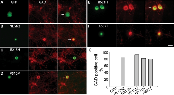
GABAergic synapse formation assay of four missense NLGN2 mutants. (A–F) Representative images showing the transfected HEK 293T cells (green) and immunostaining of GAD to visualize GABAergic terminals (Red). Cell transfected with GFP alone did not have GAD positive staining (A), while the wild-type NLGN2-expressing cell had positive GAD staining (B). Among four NLGN2 mutants (C–F), the R215H mutant did not have GAD positive staining. (G) Quantification confirms that the R215H mutant has no detectable GAD positive cells, while the other three mutants showed no differences in the counts of GAD positive cells compared the wild-type (GFP: 0 out of total 16 cells examined; wild-type NLGN2: 18/21; R215H: 0/27; V510M: 14/15; R621H: 14/17; A637T: 12/15). Arrows point to transfected HEK 293T cells in merged images. Scale bar, 10 μm.

