Fig. 8.
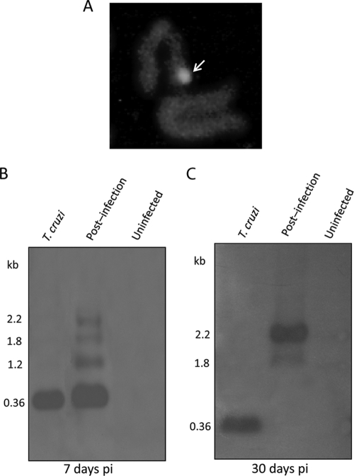
Insertion of the Trypanosoma cruzi kDNA minicircle into the human macrophage genome. (A) Identification of the kDNA in a metaphase plate chromosome by the FISH method. Fluorescence (arrow) is seen in a chromosome probed with a biotin-labeled minicircle. (Reprinted from reference 63 with permission of the publisher.) (B) Southern hybridization of NsiI digests of 7-day-postinfection (pi) macrophages with a kDNA probe on blots of 0.8% agarose gels. The T. cruzi kDNA minicircle forms a single 0.36-kb band, whereas the early-infection macrophage DNAs show upper 1.2-, 1.8-, and 2.2-kb bands. (Reprinted from reference 408 with permission of the publisher.) (C) Hybridization of NsiI digests of 30-day (postinfection) macrophages with a kDNA probe. The absence of the 0.36-kb band in the Southern blot and the presence of the 1.8- and 2.2-kb PCR products in the 30-day-postinfection macrophage DNA indicate that the T. cruzi infection is eradicated and that the minicircle is inserted into the host cell genome. (Reprinted from reference 408 with permission of the publisher.)

