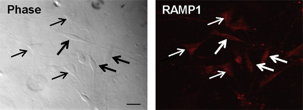
Fig. 1.
RAMP1 expression in trigeminal ganglion glia. Cultures enriched in glia were immunostained for the presence of the CGRP receptor protein RAMP1 and imaged at 400× magnification using phase contrast microscopy (phase, left panel) as well as fluorescent microscopy (RAMP1). Thick arrows indicate satellite glial cells while thin arrows indicate Schwann cells. Scale bar equals 25 µm.

