
Figure 2.
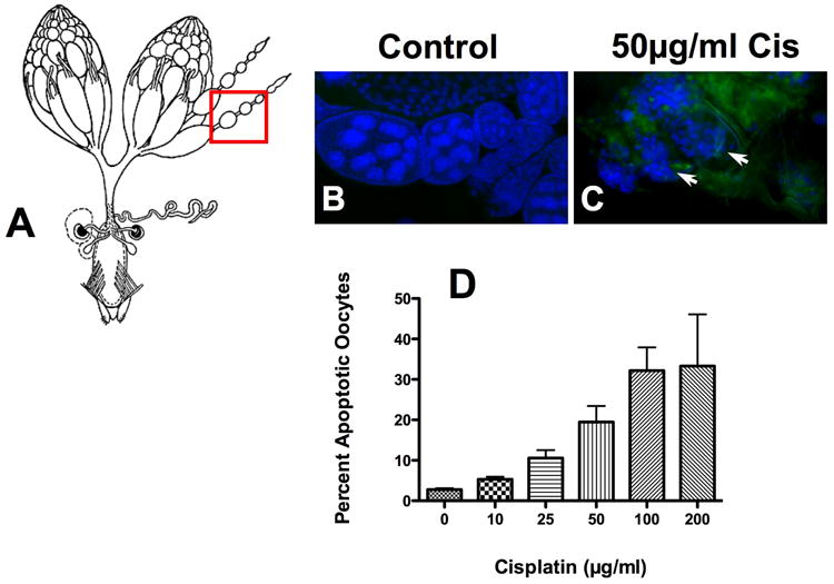
Cisplatin-induced apoptosis in Drosophila oocytes. To determine if drug delivery was effective, ovaries harvested from flies treated with and without 50μg/ml cisplatin were immunostained and oocytes were quantitated for apoptosis. (A) Ovaries (www.flybase.org/reports/FBim0000078 permissions requested) were stained for active caspase 3 and nuclei were visualized using bisbenzimide (blue). Normal oocytes with no caspase 3 activation and intact nuclei were observed in (B) controls. Fragmented nuclei (arrows) with positive caspase 3 staining (green) along with a disorganized morphology were observed in (C) cisplatin treated oocytes. (D) Caspase 3 positive oocytes were counted and expressed as a percent of the total number of oocytes (p=0.098). Despite the disorganized morphology, we were still able to count individual oocytes.