
Figure 3.
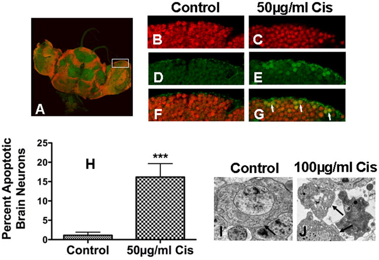
Cisplatin-induced apoptosis in Drosophila brain neurons. (A) Brain harvested from cisplatin treated (50μg/ml) flies were stained for active caspase 3 and eLav, a pan neuronal marker. Control and cisplatin treated brain neurons stained for (B, C) eLav (red) and (D, E) active caspase 3 (green), showed (F) no caspase 3 staining in control brain and (G) positive caspase 3 staining with co-localized eLav staining in cisplaitn treated brain (arrows). (H) Caspase 3 staining was found in 1.11% of control brain neurons (n=10) versus 16.18% in cisplatin treated brain neurons (n=8) (p=0.0007). Electron micrographs of (I) control brain and (J) brain treated with 100μg/ml cisplatin show apoptosis with intact plasma membrane (arrows) and fragmented nuclei.