
Figure 2. Loss of Rac1 protein and mRNA expression in a newly developed forebrain specific Rac1 mutant mouse model.
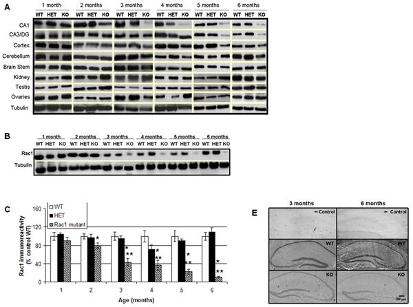

A) Representative immunoblots depicting levels of Rac1 protein in the brain and other tissues of 1-6 months old Rac1 mutant (KO) and control mice (wild type, WT and heterozygotes, HET), B) Representative E-PAGE® depicting gradual loss of Rac1 protein in the CA1 area of 1-6 months old Rac1 mutant mice (KO) relative to controls (wild type, WT and heterozygotes, HET), C) Group data (mean ± SEM, n=6) of Rac1 immunoreactivity in CA1 area of Rac1 mutant mice relative to control mice. Asterisks denote statistically significant difference respect to * wild type (WT) or ** heterozygote (HET) (p<0.05, ANOVA), D) Double staining showing localization and expression of Rac1 in hippocampal slices. Hippocampal slices were stained with anti-Rac1 (green) and DAPI (blue). Images show immunoreactive Rac1 not colocalized with DAPI (nuclear marker) and disappearing in the Rac1 mutant mice, E) In situ hybridization showing expression of Rac1 mRNA in Rac1 mutant (KO) and control animals (WT) at 3 and 6 months of age. A negative control (- Control) indicates no unspecific binding.