
Fig. 2.
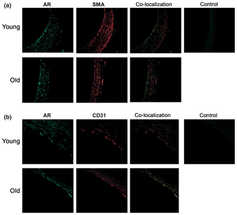
Immunostaining reveals aldose reductase (AR) expression in aortic endothelial cells and smooth muscle cells of aged animals. Formalin-embedded sections of aortas from young and aged rats were probed with anti-AR IgG, anti-alpha smooth muscle actin (SMA) (a) and anti CD31 (b) IgG, respectively, and visualized after using appropriate second antibody and fluorescent conjugated IgG. Images were visualized under 40× magnification. Immunostaining localized AR to the aortic endothelial and smooth muscle cells in aged animals. Aorta sections stained with appropriate nonimmune serum are presented as negative control.