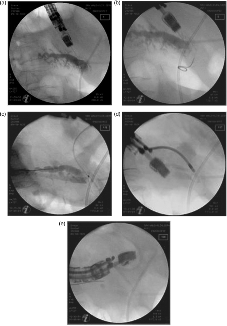
Figure 2.
Pancreaticography-based control view during the intervention. (a) EUS-guided transgastric puncture of the pancreatic duct using a 19-G needle. (b) Unsuccessful insertion of the guide wire into the pancreatic duct (attempt to direct it into the papilla of Vater from the pancreatic site). (c) Placement of the guide wire within the tail segment of the pancreatic duct. (d) Expansion of the gastropancreaticostomy using a 10-Fr retriever. (e) Placement of a 10-Fr Amsterdam prosthesis via the guide wire.

