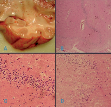
Figure 1.
Illustration of the case vignette. The Ammon’s horn was not atrophic (A). Low power magnification (B): diffuse cell loss, confirmed by large power views (C, D). No inflammation either parenchymal or around vessels was detected. (Images courtesy of Professor M Klimpfinger, Institute Pathology, KFJ Hospital, Vienna).

