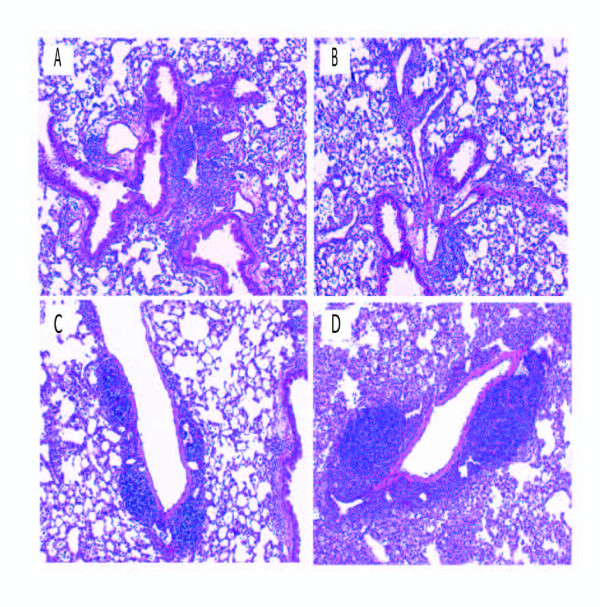
Figure 4.
Development of granulomas in lungs of WT and KO mice. Representative H & E stained lung sections from WT mice (A and C), IL-6KO mice (B), and T-Bet KO mice (D), exposed to S. rectivirgula for 3 weeks. IL-6 KO mice exposed to S. rectivirgula (B) form granulomas similar to WT mice exposed to S. rectivirgula (A). T-bet KO mice exposed to S. rectivirgula (D) demonstrated more severe granuloma formation compared to WT mice exposed to S. rectivirgula (C). (Original magnification × 63).

