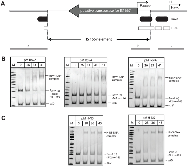
Figure 9. RovA and H-NS binding to the Y. enterocolitica O:3 invA regulatory region.
(A) Overview of the invA promoter region of Y. enterocolitica O:3 strains. The transcriptional start sites of the invA promoter and of the predicted IS1667-encoded promoter are indicated by broken arrows. The dark boxes represent the RovA and the white small boxes the H-NS binding sites identified in the homologous invA promoter of Y. pseudotuberculosis. The thick line represents the invA promoter sequence and the thin line illustrates the sequence of the IS1667 element with the putative transposase gene. Fragments used for the band shift experiments are shown as black lines. Competitive gel retardation assays using purified RovA protein (B) or purified H-NS (C) of Y. enterocolitica O:3 strain Y1. DNA fragments comprising different portions of the invA regulatory region of Y1 were incubated without or with increasing concentrations of purified RovA or H-NS. The DNA-protein complexes were separated on a 4% polyacrylamide gene, a molecular weight standard 100 bp ladder was loaded on the left. The higher molecular weight protein-DNA complexes are marked by an arrow and the positions of the non-shifted and control fragments are indicated.

