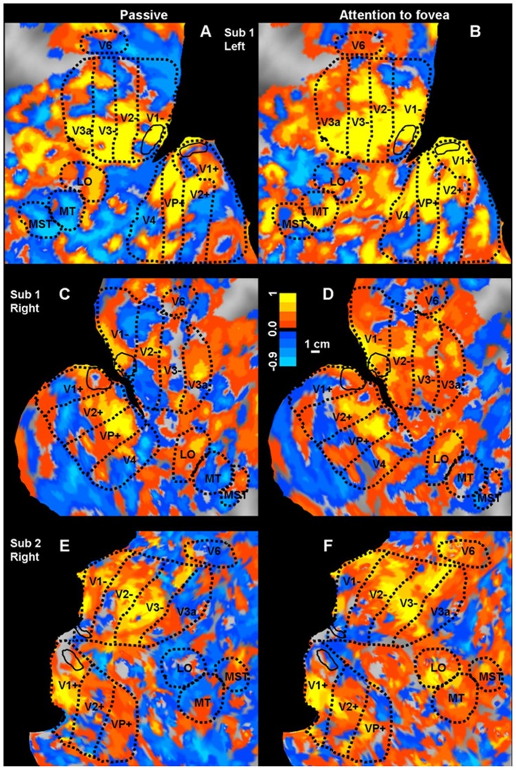
Figure 2. Gardner Spatiotopy Index of the visual cortex.
Spatiotopy index for three representative hemispheres, the left and right hemisphere of Subject 1 (A & B, C & D) and the right of Subject 2 (E & F), for the passive-fixation condition (left: A-C-E) and attention condition (right: B-D-F). During passive fixation, there are large regions of blue (spatiotopic), particularly in dorsal cortex, including areas MT, MST, V6 and, to a lesser extent, LO. However, when performing the attention-demanding foveal task areas that were clearly spatiotopic (color-coded blue) with passive fixation, become strongly retinotopic (color-coded red/yellow) when attention is directed to the fovea. The islands within V1 inside the solid black lines indicate the parafoveal regions used for the data of Figures. 8 & 1.

