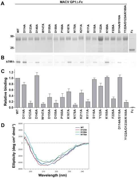
Figure 2. Binding of MACV GP1Δ variants to hTfR1 and the surface of MACV-susceptible cells.
A, expression of wild-type MACV GP1Δ, mutants thereof, and Fc control. The numbers to the left of the blots indicate relative molecular mass in kDa. B, ability of wild-type MACV GP1Δ and variants thereof to co-immunoprecipitate hTfR1. Shown is a representative western blot from two independent experiments. C, ability of these proteins to bind to the surface of MACV-permissive (Vero) cells as analyzed by flow cytometry. This assay was performed twice in duplicates yielding similar results. Bars indicate average of duplicates in one representative experiment. Results were normalized by subtracting measurements with secondary antibody only. D, far-UV circular dichroism (CD) of wt GP1Δ and mutants D123A, D159A, and N178A.

