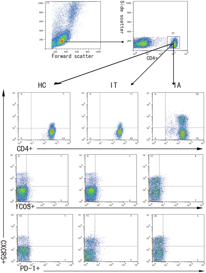
Figure 1. FACS analysis of TFH cells.
Peripheral mononuclear cells were stained in duplicate with anti-CD4, anti-ICOS, anti-PD-1 or isotype-matched IgG, respectively. The cells were gated initially on living lymphocytes (top left) and then on CD4+ T cells (top right). Subsequently, the frequency of CXCR5+CD4+, ICOS+CXCR5+CD4+, and PD-1+CXCR5+CD4+ cells were analyzed by flow cytometry. At least about 50,000 events were analyzed for each sample and data are representatives of different groups of samples from at least two independent experiments.

