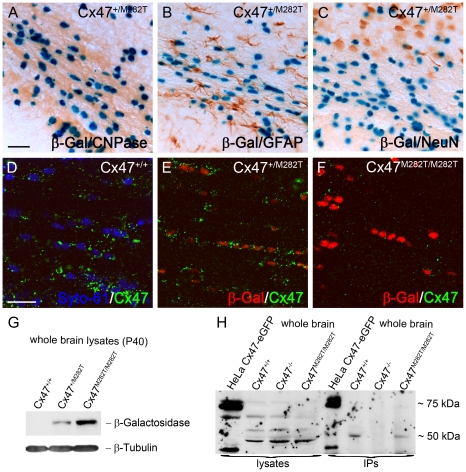
Figure 2. mCx47M282T and β-gal expression in oligodendrocytes of 40-day-old mutant mice.
A–C, LacZ staining of 50 µm brain slices obtained from Cx47+/M282T mice. The β-gal activity is restricted to nuclear localisation. A, β-Gal positive cells in the corpus callosum show typical oligodendrocytic chain-like organization and coexpression of the oligodendrocytic marker CNPase. LacZ expression was not detectable in GFAP-positive astrocytes (B) or NeuN-positive neurons (C). D, Immunostaining with Cx47 antibodies (green) revealed signals in close proximity of Syto-61 stained nuclei in the corpus callosum of Cx47+/+ mice. Weaker signals were also found more distal to the nuclei. E, Cx47 antibody stainings on heterozygous Cx47+/M282T mice resulted in weaker, but similarly localized immunosignals compared to those obtained on wildtype brain tissue. Cx47 gap junction immunosignals were predominantly localized close to β-gal positive nuclei indicated by antibody staining. F, Homozygous Cx47M282T/M282T mice showed apparent β-gal immunoreactivity, but robust Cx47 gap junction immunosignals were not detected in the perikarya. Very weak Cx47 signals were noticed in brain tissue of Cx47M282T/M282T mice. G, Immunoblot staining against β-gal on whole brain lysates of 40-day-old mice yielded strong signals with Cx47M282T/M282T, weaker signals with Cx47+/M282T and no signals with Cx47+/+ tissue. H, Immunoblot analysis on Cx47 protein resulted in unspecific bands at 50 kDa of whole brain lysates obtained from wildtype, Cx47M282T/M282T and Cx47 deleted (Cx47−/−) tissue. Immunoprecipitated Cx47 was detected in wildtype and Cx47M282T/M282T tissue. Lysates of Cx47-eGFP fusion protein expressing HeLa cells yielded signals at 75 kDa as expected and additional weaker signals at approximately 40 kDa after immunoprecipitation of Cx47 and with crude HeLa cell lysate. Additional signals at 40 kDa in these lanes may represent the cleaved C-terminal region of Cx47. Scale bars: A–C, 50 µm; D–F, 20 µm.

