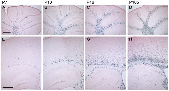
Figure 3. The number of β-gal positive cells increases during postnatal development of homozygous mCx47M282T expressing mice.
Sagittal 50 µm brain sections were X-Gal stained for LacZ expression and counterstained with eosin. The LacZ reporter gene reflects the expression of Cx47. A, In the cerebellum of 7-day-old Cx47M282T/M282T mice several X-Gal stained oligodendrocytes were already found in the medullary centre. B, Stainings on cerebella of P10 mice revealed an increase in the number of β-gal positive cells in the white matter compared to P7 mice. Single β-gal positive cells were already found in the granular layer. C and D, Cx47 expressing cells further increased in number during development of the cerebellum. In addition to their localisation in the cerebellar white matter, LacZ expressing cells were found to be widespread among the granular layer in the cerebellum of 105 day old mice. E, Compared to the number of X-Gal stained cells in the white matter of P7 cerebellum, only few cells were stained in P7 cerebral white matter, localized in the corpus callosum and the hippocampal fimbria. Single cells were found in the cortex proximal to the corpus callosum. The hippocampal gray matter was almost free of β-gal positive cells at this developmental stage. F, On P10 the number of X-Gal stained cells was remarkably increased in the corpus callosum and the hippocampal fimbria compared to P7. β-Gal positive cells decreased in number from ventral to dorsal in the corpus callosum to the cortex. G, P16 animals showed robust LacZ expression in the white matter of the cerebrum including corpus callosum, hippocampal fimbria and hippocampal alveus. Furthermore, β-gal positive cells were present at the hippocampal fissure and near the hippocampal regions Cornu ammonis 1 (CA1) and CA3. Compared to P10 the number of LacZ expressing cells increased dramatically in cortical layers VI to IV of P16 mice. H, 105 days after birth Cx47 expressing cells represented by LacZ reporter gene stainings were localized all over the telencephalon with strong accumulation and typical oligodendrocytic chain-like organizaiton in the white matter. In gray matter, β-gal positive cells were scatterly distributed with a decreasing gradient ranging from layers VI to I of the neocortex. Scale bar: A–H, 500 µm.

