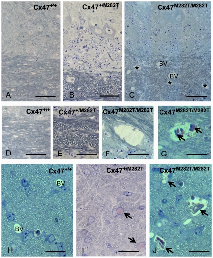
Figure 4. Toluidine blue/pyronin g stained semi-thin sections reveal cystic spaces in white matter of Cx47M282T/M282T mice.
Semi-thin sections of cerebellar white matter of Cx47+/+ (A, C) Cx47+/M282T (B, E, I) and Cx47M282T/M282T (C, F, G, J) mice are shown after staining with toluidine blue/pyronin g. Cystic spaces in white matter are indicated by asterisks in C and can be delineated from blood vessels (BV) due to their irregular lining and absence of endothelia. At higher magnification (F, G) cysts can be shown to contain cellular debris (arrows in G). No alterations were seen in cerebella from heterozygous mice (B, E). In the prechiasmatic optic fascicle of Cx47M282T/M282T mice, similar cysts are evident (arrows in J, compared to blood vessles (BV) in the WT control shown in H). Scale bars: A–C, 50 µm; D–J, 20 µm.

