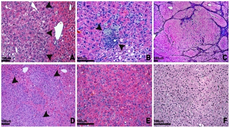
Figure 7. Evolution of steatosis to steatohepatitis and neoplasia.
(a) Histology of the liver of a 4 month-old Acox1lampe1 mouse showing fat accumulation in a zonal distribution around the central veins (magnification 200×) and (b) scattered foci of lobular inflammation with aggregates of macrophages at 4 months (magnification 400×). (c) At 12 months, the liver architecture is markedly disrupted by expansile nodules replacing most of the parenchyma (magnification 200×). (d) Foci of dysplastic hepatocytes and dysplastic nodules were present throughout the liver and some of the nodules contained areas of frank hepatocellular carcinoma (e), with loss of reticulin fibers (f). Hematoxylin and eosin (a–e) and reticulin (f) stain. 6D–F 400× magnifications.

