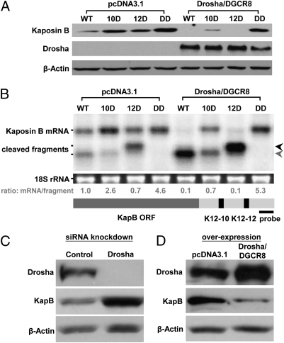
Fig. 2.
Drosha regulates KapB expression through processing of pre-miRNA cis elements. (A) Immunoblot analysis demonstrates that overexpression of Drosha and DGCR8 leads to decreased KapB expression. Various KapB expression vectors were cotransfected into HEK293T cells along with Drosha and DGCR8 expression vectors (see the legend for Fig. 1 B for description of KapB mutant vectors). Forty-eight hours after transfection, cells were harvested for immunoblot analysis. Note, at this exposure level, only overexpressed and not endogenous Drosha levels are detectable. β-Actin levels are shown as a loading control. (B) Northern blot analysis demonstrates that overexpression of Drosha and DGCR8 leads to accumulation of cleaved fragments from KapB mRNA for those constructs that retain one or more pre-miRNAs in the 3′ UTR. Transfections were conducted as described in A. A ∼220-nt radiolabeled probe complementary to the 3′ end of the KapB transcript (diagrammed at bottom of B) was used to detect full-length KapB mRNA or the cleaved products. The calculated ratio of full-length KapB mRNA to cleavage fragments is shown below each lane. Note that the fastest migrating cleavage fragment band (indicated with gray arrow) could represent the cleavage product of either premiR K12-12 alone, or cleavage of both premiR K12-12 and premiR K12-10. The black arrow indicates the fragment generated by cleavage of premiR K12-10. (C and D) Drosha levels regulate KapB expression in KSHV-infected cells. (C) Decreasing Drosha levels increases KapB expression. Drosha siRNAs were transfected into TREx-RTA BCBL-1 cells and immunoblot analysis was performed. (D) Increasing Drosha/DGCR8 levels decreases KapB expression. Drosha and DGCR8 expression vectors were cotransfected into TREx-RTA BCBL-1 cells and immunoblot analysis was performed.

