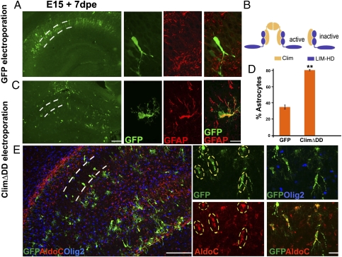
Fig. 2.
A dominant-negative construct recapitulates the Lhx2 loss-of-function phenotype. (A) WT E15 embryo electroporated with a control GFP construct displays labeled cells in a tightly packed pyramidal cell layer 7 d after electroporation (dashed lines). These cells display characteristic neuronal morphologies and do not express GFAP. Confocal images of GFP-GFAP labeling are shown alongside the low-magnification images. dpe, days postelectroporation. (B) Tetrameric model of LIM-HD function illustrating a transcriptionally active complex containing two LIM-HD molecules (blue) and two Clim molecules (yellow). A truncated Clim protein lacking the dimerization domain (ClimΔDD) blocks formation of the tetramer, and thus interferes with LIM-HD protein function. (C) Electroporation of ClimΔDD-GFP into WT embryos generates cells scattered throughout the hippocampus, which are GFAP-positive and have multipolar morphologies. Confocal images of GFP-GFAP labeling are shown alongside the low-magnification montages. (D) GFP- and GFAP-expressing cells were scored. In embryos electroporated with GFP (controls), 35% of the GFP-expressing cells were also GFAP-positive. In ClimΔDD-GFP electroporated embryos, 80% of the electroporated cells were GFAP-positive. The bars represent the mean ± SD (**P < 0.001). (E) ClimΔDD-GFP electroporated cells (green cells marked by dashed ovals) express astrocyte-specific marker AldoC (red) but do not express oligodendrocyte marker Olig2 (blue). (Scale bars: low-magnification images, 100 μm; high-magnification images, 20 μm.)

