Fig. 3.
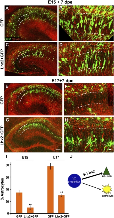
Lhx2 overexpression enhances and prolongs neurogenesis. (A and B) WT embryo electroporated with a control GFP construct displays several labeled cells in the pyramidal cell layer 7 d after electroporation (dashed lines). These cells display characteristic neuronal morphologies and do not express GFAP. Other cells are scattered in extrapyramidal locations, and several of them coexpress GFAP. dpe, days postelectroporation. (C and D) Electroporation of a full-length Lhx2 construct enhances neurogenesis, with fewer cells occupying extrapyramidal positions and expressing GFAP. (E and F) WT E17 embryo electroporated with a control GFP construct displays labeled cells scattered in extrapyramidal locations 7 d after electroporation (dashed lines). These cells display characteristic astroglial morphologies and coexpress GFAP. The pyramidal layer (dashed lines) appears nearly devoid of GFP cells. (G and H) Electroporation of a full-length Lhx2 construct enhances neurogenesis, with several cells occupying the pyramidal cell layer in a tight band. These cells display neuronal morphologies and do not coexpress GFAP. (I) GFP- and GFAP-expressing cells were scored in electroporated brains. For E15 electroporations, the proportion of GFP cells that were also GFAP-positive in control GFP embryos was 35%, and in Lhx2-overexpressing embryos, the proportion was 10%. In control E17 embryos electroporated with GFP, the proportion was 79%, and in Lhx2-overexpressing embryos, the proportion was 31%. The bars represent the mean ± SD (**P < 0.0001). (J) Diagram illustrating that Lhx2 overexpression enhances neurogenesis. All high-magnification images are generated from montages of confocal images of GFP (green) and GFAP (red). (Scale bars: low-magnification images, 100 μm; high-magnification images, 20 μm.) A–H are composites assembled from multiple confocal images.

