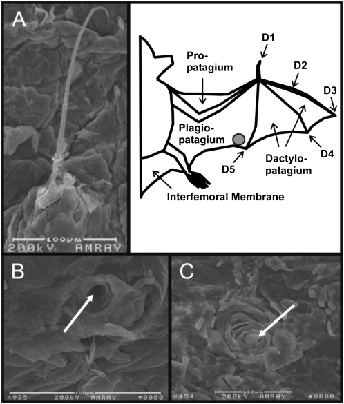
Fig. 1.
Sensory wing hair before (A) and after (B and C) depilation. (A) Scanning electron microscope image from a domed hair located on the ventral trailing edge (location is marked by a gray circle in schematic to the Right) of Eptesicus fuscus. Note the calibration bar below the photomicrograph for reference. (Right) Schematic of the bat wing and its parts. (B and C) Examples of domes after depilation. Arrows point to the center of the domes from which the hair would normally protrude.

