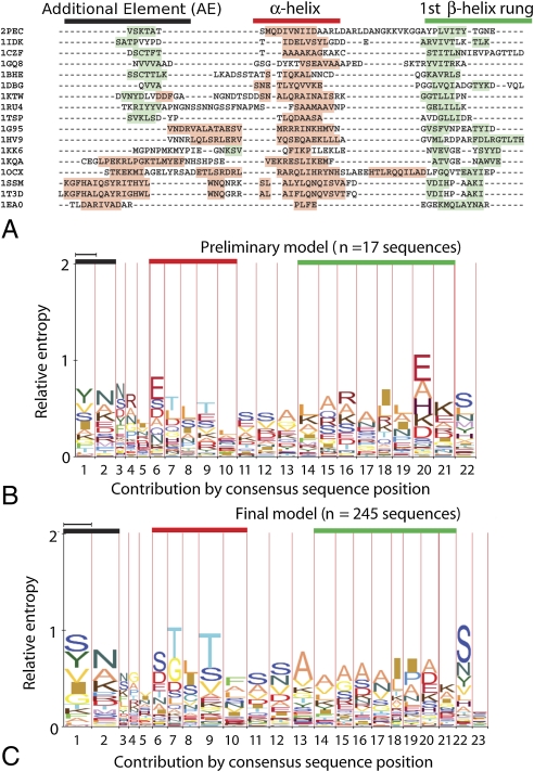
Fig. 3.
Alignment and prediction of α-helix caps. Black bars denote the AE, red the α-helix, and green the first rung of the β-helix. (A) Structurally based alignment. At top, single α-helix caps from the right-handed β-helix pectate lyase superfamily; at bottom, single and double α-helix caps from the left-handed β-helix superfamily. Shading denotes secondary structure by PDB annotation: pink, α-helix; light green, β-strand. (B, C) HMM-Logo representations (18) of the α-helix-cap predictive model. Narrow-column positions are more likely to align with gaps than wide columns. (B) The initial model constructed from 26 aligned crystal structures in 17 families. (C) The augmented model constructed from 1,084 sequences aligned to the initial model.

