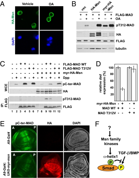
Fig. 5.
Msn is activated on membrane translocation and inhibits Dpp signaling in vivo. (A) Immunofluorescence staining showing the plasma membrane translocation of HA-Msn in S2 cells after OA (125 nM, 1 h) treatment. (B) HA-Msn with or without a myristoylation sequence was coexpressed with FLAG-MAD in S2 cells. FLAG-MAD was immunoprecipitated, and the level of T312 phosphorylation was examined. The effect of OA treatment is shown where indicated. (C) S2 cells were transiently transfected with indicated combinations of plasmids. After Dpp treatment, the whole cell extract (WCE), or the FLAG proteins (after IP) were analyzed using the indicated immunoblots. (D) The same cells from lanes 4, 6, 10, and 12 in (C) were analyzed by quantitative real-time PCR to measure the mRNA level of dad, using Rp49 as the standard (mean ± SD). (E) Third instar wing imaginal discs from the indicated transgenic flies were double-immunostained with pC-ter-MAD (green) and HA (red). The nuclei were stained by DAPI (blue). The orientation of the wing disk is denoted by “A” for anterior and “P” for posterior. (F) A model showing direct regulation of Smad by Msn kinases. The activity of Msn is regulated by as-yet unknown mechanisms.

