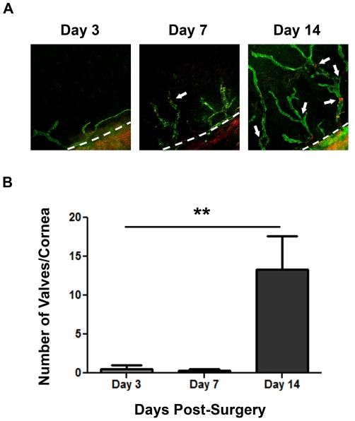
Figure 3. Time course of lymphatic valve formation during corneal inflammation.
(A) Representative micrographs demonstrating the increase of lymphatic valves in sutured corneas with the progression of corneal inflammatory lymphangiogenesis. Itga-9: red; LYVE-1: green. Original magnification: 50 X. Dashed lines: demarcation between the cornea and conjunctiva. (B) Summarized data from repetitive experiments showing lymphatic valve quantification at day 3, 7, and 14 after suture placement. **P<0.01.

