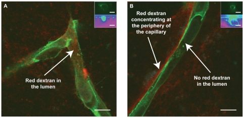
Figure 3. Dextran tracer localization via confocal microscopy.
60x confocal microscopy images of days 5 (A) and 10 (B) capillaries after 70 kDa dextran tracer addition (red), fixation with 10% formalin, and human CD31 staining (green) of HUVECs. Inset images: HUVECs (top) and Dextran (bottom) show hollow lumens with or without dextran. A) Red tracer is present within the lumens of the capillaries, demonstrating a low resistance to permeability. B) Red tracer is excluded from the lumens of the capillaries, demonstrating an increased resistance to permeability as the capillaries mature over time.

