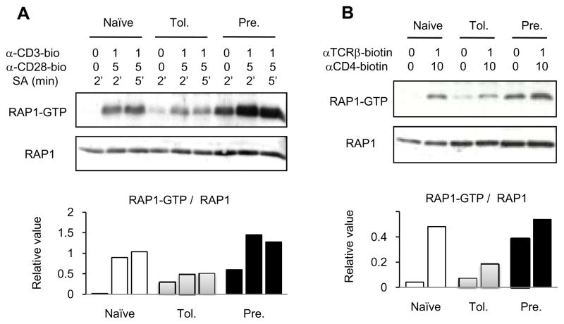
Figure 2. RAP1 activation in adaptively tolerant T cells.
A and B, Purified naïve, adaptively tolerant, or pre-activated T cells were unstimulated or stimulated for 2 or 5 min with anti-CD3-biotin mAb (1μg/ml) and anti-CD28-biotin mAb (5μg/ml) (A) or for 1 min with anti-TCRβ-biotin mAb (1μg/ml) and anti-CD4-biotin mAb (10 μg/ml) (B), followed by cross-linking with streptavidin. Samples were lysed with 1% NP40 buffer, and active GTP-bound RAP1 was detected with a pull-down assay using GST-fusion RAL GDS-RBD. Bound RAP1 (upper gels) and total RAP1 (lower gels) were detected by Western blotting with anti-RAP1 Ab. The density of each band was determined using GelPro software. The relative values normalized to the total level of RAP1 expression are shown in the lower figures. The data show one representative experiment from two that were performed.

