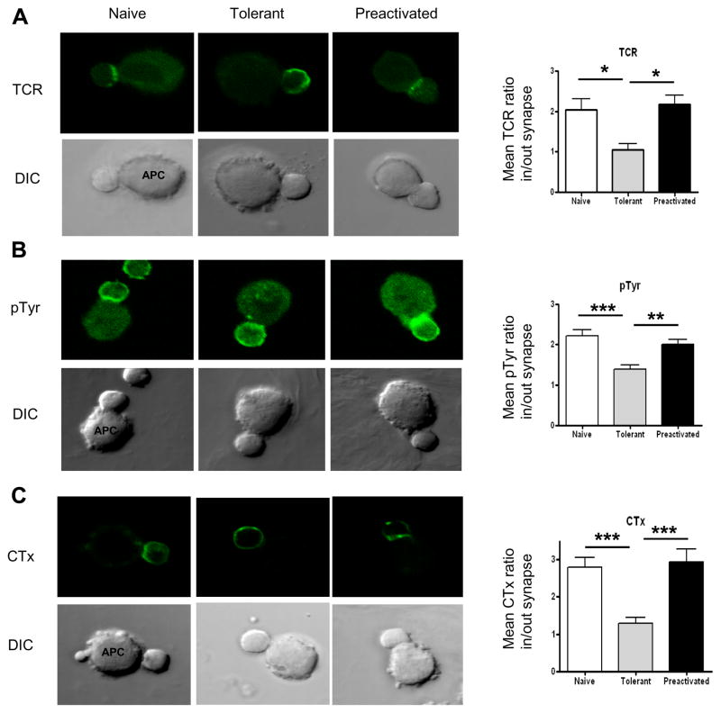
Figure 5. Translocation of the TCR, tyrosine-phosphorylated proteins, and lipid rafts to the T cell/APC contact site is impaired in adaptively tolerant T cells.
A and B, Purified naïve, adaptively tolerant or pre-activated T cells were stimulated for 5 min with the P13.9 cell line pre-pulsed with 1μM MCC (88–103). Cells were fixed, permeabilized, and stained for TCRs or tyrosine phosphorylated molecules. Representative images for Tcell/APC conjugates and the localization of TCRs or tyrosine phosphorylated molecules are shown in the left panels. The ratios of the mean number of phosphorylated molecules or TCRs in/out of the synapse were measured with ImageJ software and are shown in the right panels. *, p < 0.05; **, p < 0.01; ***, p < 0.001.
C, Purified naïve, adaptively tolerant, or pre-activated T cells were stained with FITC- CTx and then incubated with the P13.9 cell line pre-pulsed with 1μM MCC (88–103). After 5 min, the cells were fixed. In the left panels, representative images for Tcell/APC conjugates and localization of the lipid rafts are shown. The mean CTx ratios in/out of the synapse were measured with ImageJ software and are shown in the right panel. Thirty conjugates were counted in each of 2 experiments. ***, p < 0.001.

