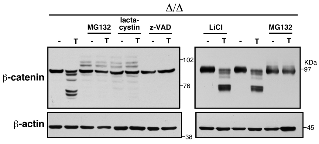
Fig. 3.
tak1 Δ/Δ keratinocytes were pretreated with the indicated inhibitors for 0.5 h (LiCl) or 1.0 h (other inhibitors) and stimulated with 20 ng/ml TNF for 4 h (left panels) or 4.5 h (right panels). Cell lysates were immunoblotted with anti-β-catenin. 10 µM MG132, 5 µM lactacystin, 20 µM Z-VAD-fmk (Z-VAD), and 10 mM LiCl were used.

