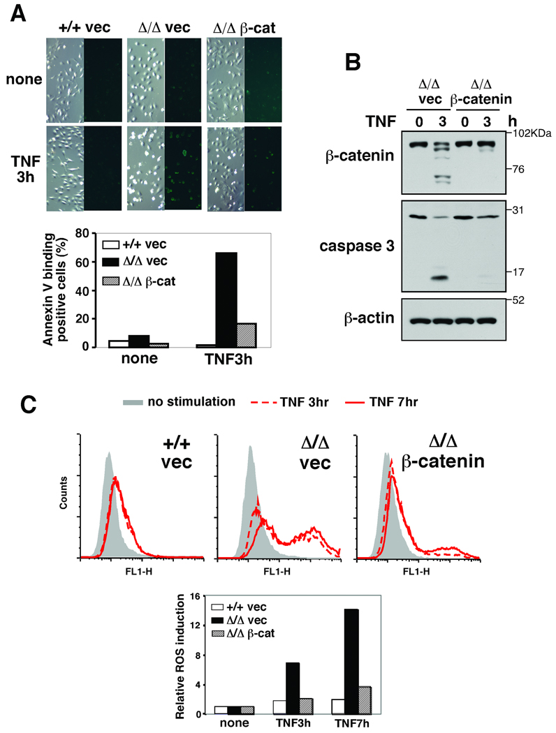
Fig. 4.
(A) tak1 +/+ and Δ/Δ keratinocytes expressing vector or β-catenin were stimulated with 20 ng/ml TNF for 3 h and apoptotic cells were stained by Annexin V-Alexa Fluor 488. (B) tak1 Δ/Δ keratinocytes expressing vector or β-catenin were stimulated with 20 ng/ml TNF for 3 h and cell lysates were immunoblotted with the indicated antibodies. (C) tak1 +/+ and Δ/Δ keratinocytes expressing vector or β-catenin were stimulated with 20 ng/ml TNF for 3 or 7 h. Cells were subsequently incubated with 10 µM CM-H2DCFDA for 30 min and analyzed by flow cytometry. The fluorescence units relative to that of unstimulated cells are also shown.

