Figure 5.
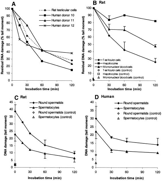
Induction and repair of DNA lesions in different cells exposed to MMS. DNA damage (tail moment; mean ± SD) was measured with the Comet assay. (A) Repair of MMS-induced DNA lesions in human and rat male germ cells. Rat testicular cells were exposed to 0.2 mM MMS, whereas human testicular cells (donors 10–12) were exposed to 0.2, 0.6 and 0.8 mM MMS, respectively, and incubated for up to 2 h to allow repair. The initial tail moment is defined as 100%. Broken line with diamonds shows rat testicular cells; solid lines with squares, triangles and crosses are human testicular cells from human donors 10, 11 and 12, respectively. (B) Repair of MMS-induced DNA lesions in testicular compared to somatic cells. Rat testicular cells, MNCs and hepatocytes were treated with 0.2, 0.5 and 0.5 mM MMS, respectively and analysed as in (A). Diamonds, squares and triangles represent testicular cells, hepatocytes and MNCs, respectively. Filled symbols show exposed cells; open symbols show non-exposed cells (controls). (C) Repair of MMS-induced DNA lesions in rat male germ cell of different spermatogenic stages. Rat testicular cells were exposed to 0.2 mM MMS, analysed as in (A) and grouped according to ploidy: haploid, round spermatids; predominantly tetraploid primary spermatocytes. Diamonds and triangles represent round spermatids and primary spermatocytes, respectively. Filled symbols show exposed cells; open symbols show non-exposed cells (control). (D) Repair of MMS-induced DNA lesions in different human spermatogenic stages. Conditions were similar as in (C) except that the cells were exposed to 0.8 mM MMS. One representative experiment out of three is shown.

