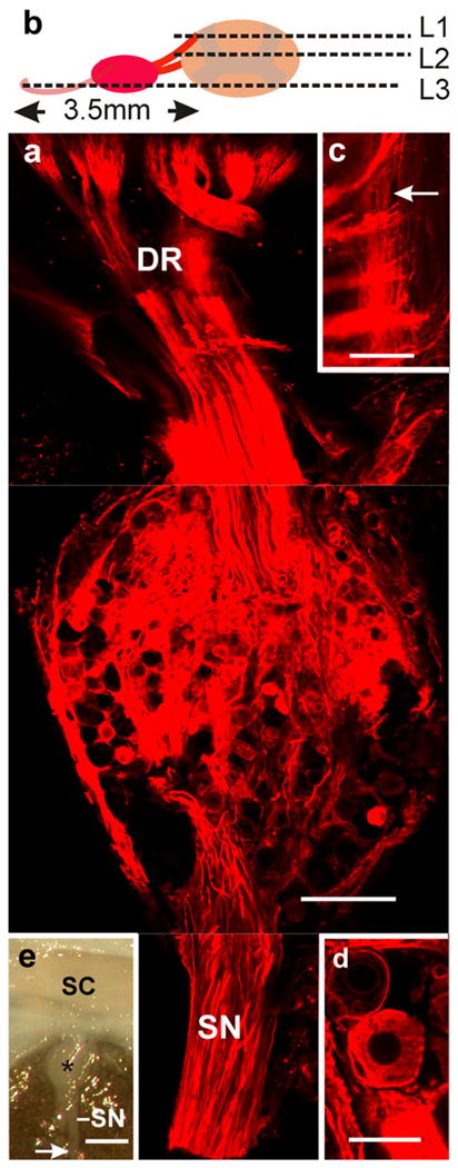
Fig. 1.

Transganglionic labeling of primary sensory fibers. a Whole ganglion combining, within a single plane, confocal optical sections taken at levels 1–3 (L1–L3) as indicated in b (SN sensory nerves, DR dorsal roots). c High magnification view of level 1 illustrating the penetration of dorsal roots into the cord and the distribution of the thinner longitudinally running branches of the sensory axons (arrow). d Note that the lipophilic dye stains the neural components of the ganglion without spreading into the extracellular space. e Macro-image depicting the arrival of the main sensory nerves (SN) to a dorsal ganglion (star). The arrow indicates the site at which the nerves were cut and DiI crystals applied with a micropipette without spurious contamination of the cord surface (enclosed in the vertebral canal; SC spinal cord). Bars 250 μm (a), 100 μm (c), 25 μm (d), 1 mm (e)
