Fig. 3.

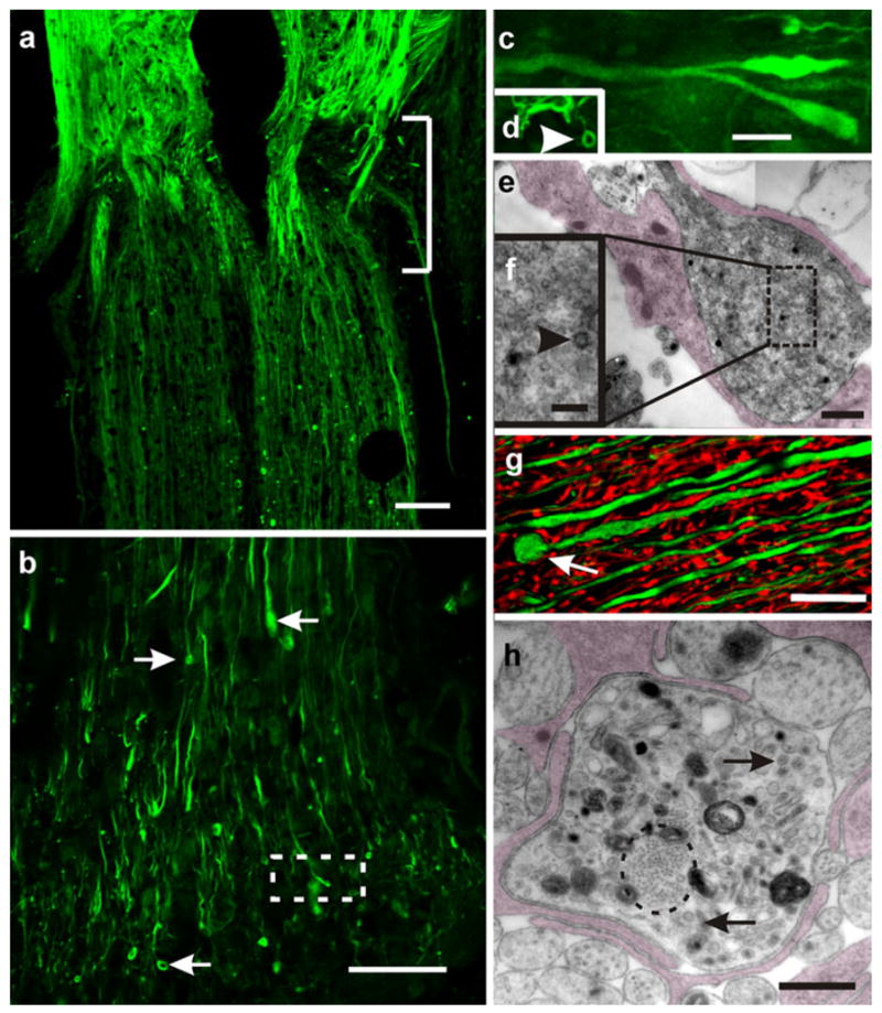
Regenerating axons cross the injured site. a Longitudinal section of a transected cord 20 days after injury. Regenerating axons stained for Neurofilament M (NF-M; green) can be seen crossing the injured site (bracket) and invading the caudal stump; confocal optical section. b Enlarged view of a bundle of regenerating axons that have penetrated the caudal portion of the cord (small arrows enlarged tips and the ring-like endings of regenerating axons); confocal optical section. c Area within the box in b examined at higher magnification. Note the presence of lanceolated and club-like “growth cones” intermingled with small terminal “rings” (arrowhead in d); confocal optical section). e Zone caudal to the injured site. Enlarged axon profiles identified as growth cones containing clusters of small vesicles; collage of two images obtained by transmission electron microscopy (TEM). The growth cone appears partially covered by glial processes (shadowed). The box is shown in f at a higher magnification. Note the presence of a characteristic “coated vesicle” (arrowhead). g “Retraction balls” or dystrophic end bulbs appear spherical (arrow) and are usually larger than the growth cones; stack of five confocal optical sections. Double-immunostaining with NF-M (green) and glial fibrillary acidic protein (GFAP; red). h TEM image showing a cross section passing through a dystrophic end bulb. Note the presence of a bundle of neurofibrils (encircled), mitochondria, cumuli of large vesicles (arrows), and dense bodies with the aspect of phagosomes. The ending is partially covered by glial lamellae (shadowed). Bars 200 μm (a), 100 μm (b), 5 μm (c, d), 0.5 μm (e), 100 nm (f), 20 μm (g), 1 μm (h)
