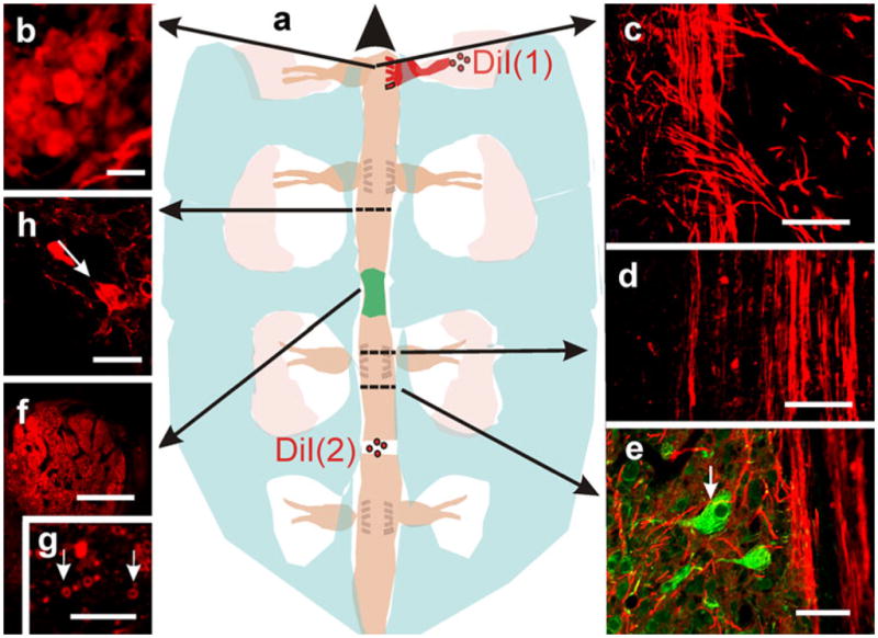
Fig. 4.

Origin of regenerating axons. a Representation depicting two different experiments with DiI as an axonal marker (long arrows correlation of the topology of the microscopic images with the gross anatomy of the cord; large arrowhead rostral). In one experiment (DiI (1)), crystals of the dye were applied to sectioned sensory nerves entering into a dorsal root ganglion at one side of the injured cord and lying cephalic with respect to the lesion epicenter (shadowed green). b Stained neurons in the ganglion (epifluorescence microscopy). c Sensory axons entering the dorsal horns. d, e Confocal optical sections revealing the occurrence of stained axons that have crossed the lesion site and have ramified close to neuronal bodies (arrow in e; DiI+NF-M). f When dye crystals were applied to the cut surface of the cord, caudal to the lesion site, conspicuous bundles of stained axons were detected at the bridge level; confocal optical section. g Higher magnification view of myelinated axons crossing the bridge; confocal optical section. h The parent neurons of some of the crossing axons (arrow); confocal optical section. Bars 10 μm (b, f, g), 30 μm (d), 40 μm (e), 50 μm (h), 100 μm (c)
