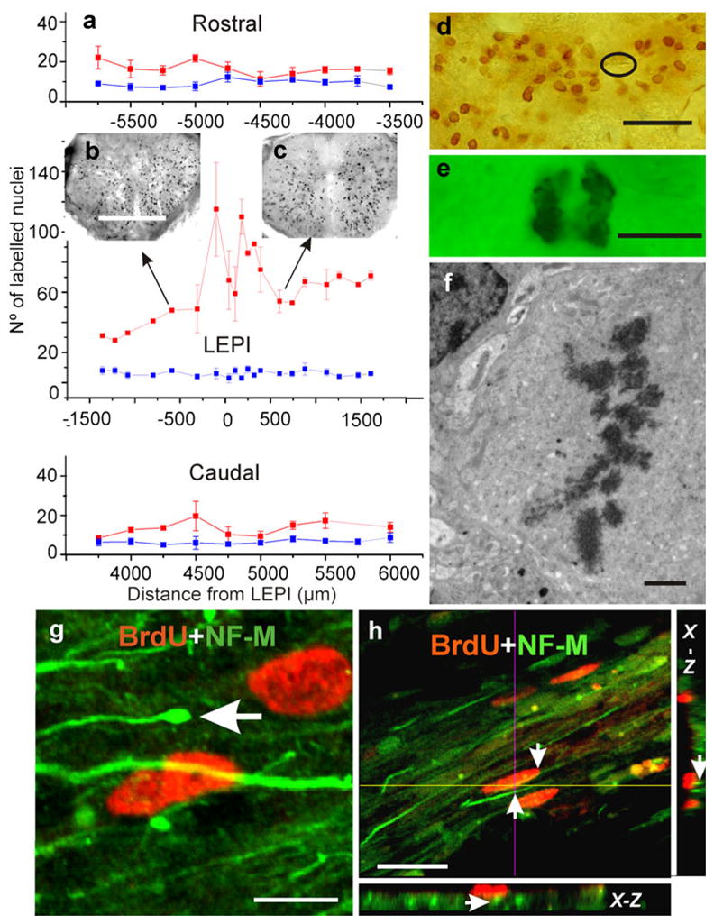
Fig. 7.

Spinal cord injury increases cell proliferation. a Mean number of BrdU-positive nuclei ± the standard deviation plotted against distance (in μm) from the lesion epicenter (red line, LEPI) and corresponding segment in control animals (blue line). Note that this is a single plot divided into three sequential rostro-caudal portions (negative numbers progression toward more cephalic segments). b, c Two small spinal cord images showing the distribution of BrdU-positive nuclei at two levels of the cord (arrows). d Even though BrdU-positive nuclei were found in all areas of the cord, they appeared concentrated near the reduced central canal (encircled). e Mitotic figures were usually found together with BrdU-positive nuclei; transmission light microscopy. f TEM image of cell in meta-phase; injured but not BrdU-injected material. g, h Double-labeling experiments revealed a close association between cells with BrdU-positive nuclei and the bundles of regenerating axons that expressed NF-M and that crossed the lesion site (arrow in g enlarged distal portion of a regenerating axon). h Proximity of two NF-M-positive axons to a BrdU-positive nucleus (arrows in h); planes obtained across a stack of 20 optical sections (x-z orthogonal plane). Bars 300 μm (b), 30 μm (d), 5 μm (e), 2 μm (f), 5 μm (g), 20 μm (h)
