Fig. 8.

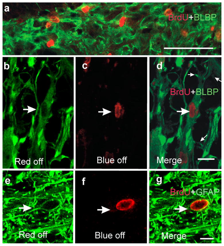
Cycling cells express glial markers. a Low power confocal microscope image (stack of five optical sections) showing several brain lipid-binding protein (BLBP)-expressing cells with their nuclei labeled with BrdU (note the random cellular organization). b–d High magnification images demonstrating that some cells expressing BLBP have BrdU-labeled nuclei (large arrows). d Merged image (small arrows main processes arising from the elongated cell body); confocal optical section. e–g Immunohistochemistry against GFAP showing abundant fibers surrounding a BrdU-positive nucleus; confocal optical section. g Merged image showing that the BrdU-positive nucleus pertains to a GFAP-positive cell. Bars (only shown in a, d, g) 50 μm (a), 10 μm (b–g)
