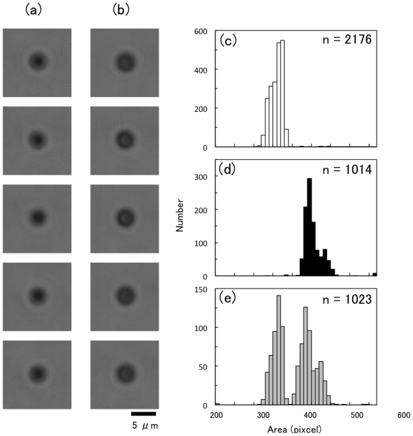
Figure 9.
Real-time size-discrimination of two similar sized particles by microscopic image. (a and b) Bright-field microscopic images of polystyrene particles with 2.5 μm (a) and 3.0 μm (b) in diameter. (c–e) Flow rate was 10 mm/s. Histograms of particles’ area measured by the imaging cytometry system. Sample suspension containing only 2.5 μm particles (c), only 3.0 μm particles (d), and mixed suspension with 2.5 and 3.0 μm particles (e).

