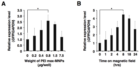
Figure 4.
Optimum conditions for PEI max-MNPs magnetofection. To optimize conditions, we varied volume (A) and time on the magnetic plate (B). These results were evaluated by quantitative real-time RT-PCR. The relative expression level (GFP/GAPDH) in the human fetal lung-derived fibroblasts (TIG-1 cells) treated with PEI max alone (A), and in the absence of magnetic force (0 h) (B) was defined as 1. Optimal transfection conditions were established when TIG-1 cells were treated with 0.8 μg PEI max-MNPs and 2.0 μg pCAG-GFP for 8 h on the magnetic plate in either a six-well plate or a 35 mm dish. The asterisk (*) indicates a significant difference (P < 0.05).

