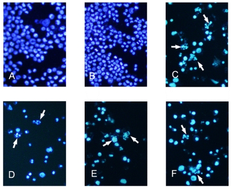
Figure 3.
Fluorescent staining of nuclei in tryptanthrin-treated K562 cells by Hoechst 33258. Cells were incubated in the medium without tryptanthrin (A) or with 0.5% DMSO (B), CTX (C) and with 6.25, 12.5, 25 μg/mL tryptanthrin (D, E and F) for 48 h, respectively. Fragmented or condensed nuclei could be observed at 200× magnification in the tryptanthrin-treated group as indicated by the arrows.

