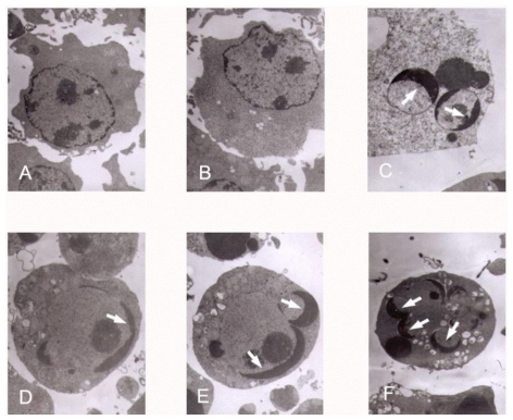
Figure 4.
Transmission electron micrographs of K562 cells treated with different concentrations of tryptanthrin. No abnormal changes were observed in the control (A) and 0.5% DMSO (B) groups. Pyknosis, chromatin margination and the formation of apoptotic bodies (white arrows) were clearly observed in the presence of CTX (C) and tryptanthrin at the concentrations of 6.25 (D), 12.5 (E) and 25 (F) μg/mL, respectively.

