
Figure 3.
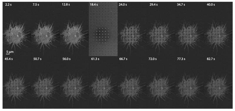
FRAP experiment with the PAM. A431 cells expressing mCitrine-erbB3 were photobleached with a dot array pattern for 6 s, followed by 60 s of imaging. Relatively long photobleaching times were required in this particular case because of the relatively low laser power available. Images were acquired at ~3 s intervals. Bar, 5 μm. Photobleaching occurred 18.4 sec into the experiment. The images have been contrast-stretched for presentation purposes.