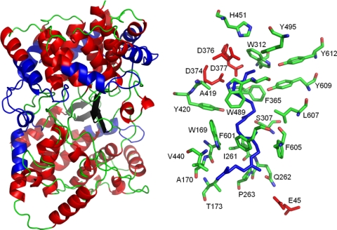
Fig. 2.
Structure and active site of the A. acidocaldarius SHC. Dumbbell-shaped structure of chain A (left) with a more structured α-barrel structure in domain 1 (upper part). The α-helices are shown in red, β-sheets in black, and loops in green. The QW motifs in the junction of helices and loops are in blue. Amino acids (sticks) of the active site together with substrate analogue 2-azasqualene (blue) are shown on the right. The aspartate residues of the DXDD motif important for the protonation initiated reaction are shown in red, as well as the glutamate residue 45, responsible for the final deprotonation. Images were generated on the basis of pDB entry 1ump by Pymol program, version 0.99rc6.

