Fig. 3.
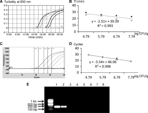
Quantitative detection of Salmonella enterica serovar Typhimurium LT2 in spiked cantaloupe samples by PMA-LAMP and PMA-qPCR. Three sets of independent spiking experiments were performed, and the LAMP reactions were repeated two times for each set of inoculations. (A) Representative PMA-LAMP amplification graph. (B) Standard curve generated for PMA-LAMP. (C) Representative PMA-qPCR optical graph. (D) Standard curve generated for PMA-qPCR. (E) Representative gel image generated by PMA-PCR. Samples 1 to 7 correspond to spiked cantaloupe samples containing 10-fold serially diluted viable Salmonella cells ranging from 6.1 × 10⁷ to 61 CFU/g in the background of dead Salmonella at 4.2 × 10⁶ CFU/g; sample 8 is water.

