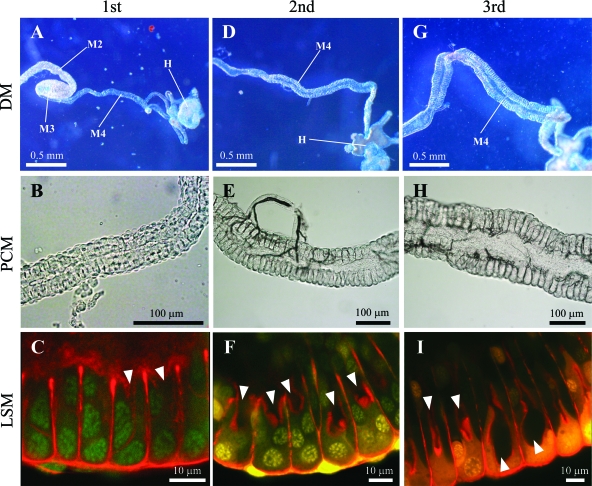
Fig. 5.
Development of midgut symbiotic organs in uninfected nymphs of Riptortus pedestris. (A to C) 1st instar nymph; (D to F) 2nd instar nymph; (G to I) 3rd instar nymph. Dissection microscopic (DM), phase-contrast microscopic (PCM), and laser-scanning microscopic (LSM) images are shown. In the LSM images, nuclei and cytoskeleton underlining the cell membrane were visualized in green/yellow and red, respectively. Arrowheads indicate the lumen of the midgut crypts. Abbreviations: M2, midgut 2nd section; M3, midgut 3rd section; M4, midgut 4th section with crypts (symbiotic organ); H, hindgut.

