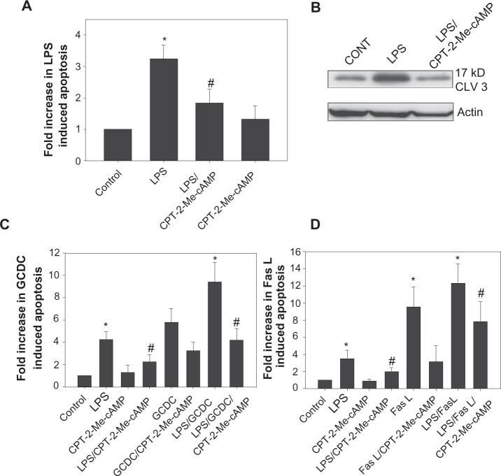
Figure 4.
Activation of cAMP-GEFs attenuates LPs induced hepatocyte apoptosis. Hepatocytes were pretreated with 20 μM cPT2-Me-cAMP or vehicle for 30 minutes followed by treatment with vehicle or 500 ng/mL LPS overnight. Cells were evaluated morphologically by Hoechst staining for the presence of apoptosis (A). Results are expressed as a percentage of the amount of apoptosis seen in the control and represent mean ± SD (n = 4–6). Whole cell lysates were immunoblotted for the 17 kD cleaved fragment of caspase 3 and equal protein loading was verified by immunoblotting for actin (B). Hepatocytes were pretreated with 20 uM cPT-2-Me-cAMP for 30 minutes and then treated overnight with 500 ng/mL LPS ± pretreatment before exposing to 100 μM GCDC for 2 hours (C) or 50 ng/mL of Fas L for 4 hours (D). *Significantly different from control. #Significantly different from LPS, P < 0.05.
Abbreviations: LPS, lipopolysaccharide; cAMP, cyclic adenosine monophosphate; CPT-2-Me-cAMP, 4-(4-chlorophenylthio)-2’-O-methyladenosine-3’5’cyclic monophosphate; GCDC, glycochenodoxycholate; SD, standard deviation.

