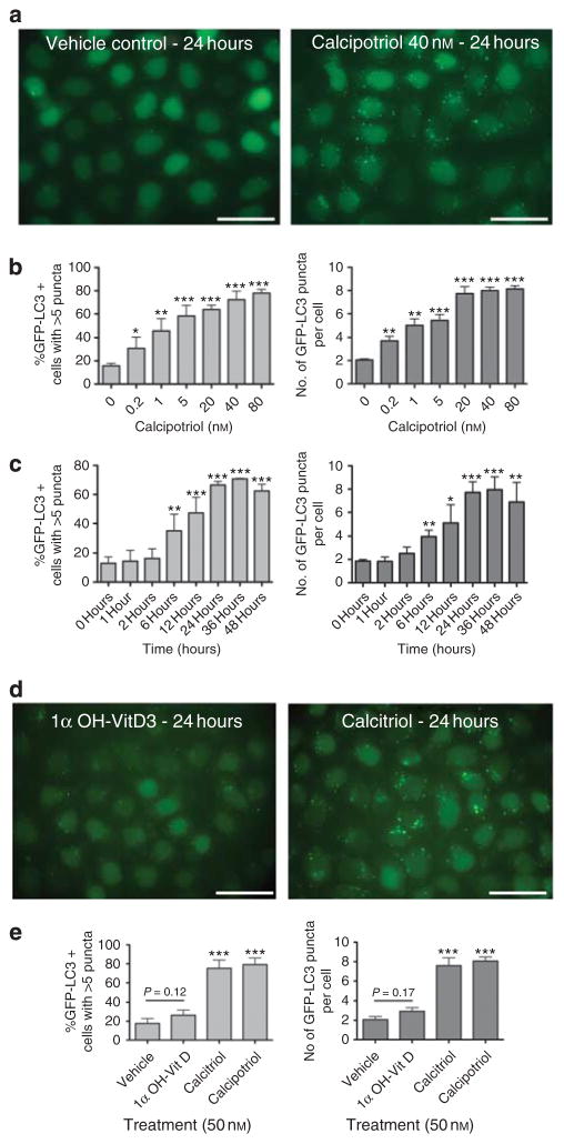
Figure 1. Calcipotriol-induced accumulation of GFP-LC3 puncta (autophagosomes).
(a) HeLa-GFP-LC3 cells were treated with 40 nM calcipotriol (Sigma, St Louis, MO) or ethanol vehicle for 24 hours. Cells were fixed in 3% paraformaldehyde (PFA) and imaged. (b, c) Effects of (b) doses and (c) durations of calcipotriol treatment on GFP-LC3 puncta. For dose titration, cells were treated for 24 hours. For time course, cells were treated with 40 nM calcipotriol. Bar graphs show percentage of cells with >5 puncta (light gray) and average number of puncta per cell (dark gray). (c) Effects of calcitriol (Cayman Chemical, Ann Arbor, MI) or 1α-hydroxyvitamin D3 (Sigma) on GFP-LC3 accumulation in HeLa-GFP-LC3 cells. (d) Quantitation of effects of vitamin D analogs. In b, c, and e, >100 cells were scored for each sample. Results represent mean±SD for triplicate samples. Similar results observed in three independent experiments. *P<0.05, **P<0.01, ***P<0.001 versus control condition; Student’s t-test. Scale bar = 50μm.

