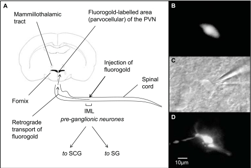
Fig. (4).
Methods for patch-clamping retrogradely-labelled neurones. A, the retrograde tracer fluorogold is injected into the rat intermediolateralis (IML) at level T2-T4, it is also possible to use other tracers, such as rhodamine-labelled microspheres (see Fig. 8). The IML is dense with pre-ganglionic neurones that project to the superiocervical (SCG) and stellate (SG) ganglia, and from there to the heart and blood vessels [65, 227]. The appearance of a fluorogold-labelled neurone B, prior to patch clamp recording, C, during patch-clamp, under near infrared differential interference contrast microscopy, and D, when patched with Lucifer yellow (a fluorescent dye) in the patch clamp pipette. The dye fills the neurone, and this gives re-confirmation that recording was from the appropriate cell. Reproduced from [43], with permission.

