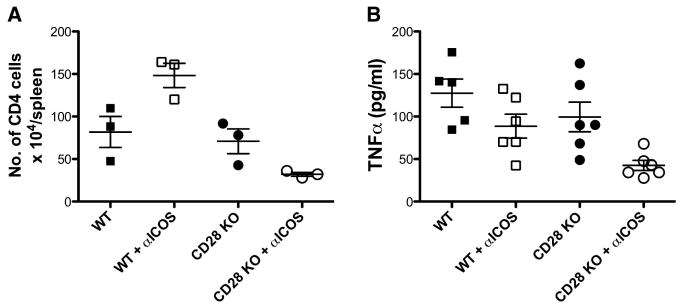
Fig. 3. Role of CD28, CTLA4 and ICOS in GVHD induced by CD4+ T cells.
Lethally irradiated BALB/c mice were transplanted with TCD-BM from normal B6 Ly5.1+ mice or plus purified CD4+ cells from WT or CD28-/- B6 donors. Half of the recipients were also treated with anti-ICOS or control mAb. (A) Six days after BMT, recipient spleen was collected and stained for expression of CD4, Ly5.1 and H2b. Data show absolute number of donor T cells (CD4+Ly5.1- H2b+) in individual mouse (n = 3 in each group), which represents one of 2 replicate experiments with similar setting. (B) In separate experiments as described in A, recipient peripheral blood samples were collected 3 weeks after BMT. The level of TNFα in recipient serum was shown in individual mouse (n = 5 or 6 per group), and the data were pooled from 2 replicate experiments.

