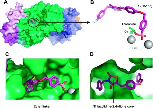
Figure 1.
ATX structure liganded with inhibitor 1 (PDB ID 2XRG). (A) Surface representation of ATX with inhibitor 1 (magenta). (B) Binding of inhibitor 1 to the threonine oxygen nucleophile and two zinc ions. (C) Visualizing the ether linker of inhibitor 1 bound to ATX. (D) Visualizing the degree of freedom for the thiazolidine-2,4-dione core of inhibitor 1 in the ATX binding site.

