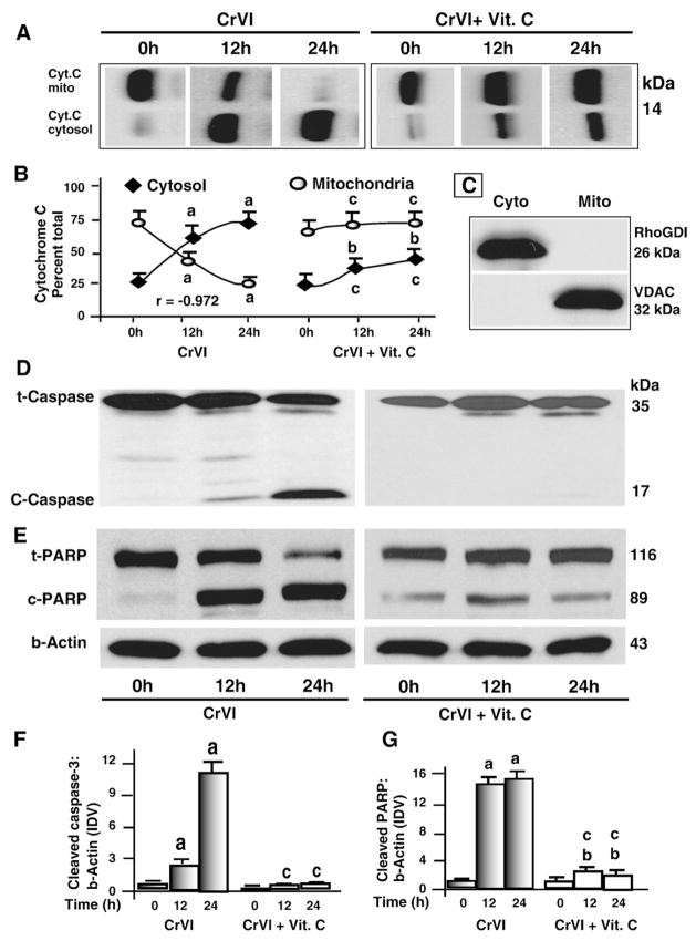
Fig. 2.
Time-course effect of CrVI on cytochrome c release from mitochondria, cleavage of caspase-3 and PARP in granulosa cells. Cells were pre-treated with or without vitamin C for 24 h and treated with CrVI (10 μM) for 0 h, 12 h and 24 h period. Protein (50 μg of either total protein or mitochondrial or cytosolic fraction) from each sample was subjected to western blot analysis as described in Materials and methods. A, Western blots of cytochrome c; B, Quantification of cytochrome c in cytosol and mitochondria with time; C, Western blots of mitochondria-specific voltage-dependent anion channel (VDAC) and cytosol-specific RhoGDI in mitochondrial and cytosolic protein fractions. Western blots of D total (t) and cleaved (c) caspase-3; and E total (t) and cleaved (c) PARP, representative blots are shown. Quantification of average protein levels of F caspase-3 and G PARP from three individual experiments is shown. Each value is the mean±SEM of three experiments, P<0.05; a: CrVI-treatment, 0 h vs 12 h or 24 h; b: CrVI+Vitamin C-treatment, 0 h vs 12 h or 24 h; c: CrVI (0 h or 12 h or 24 h) vs CrVI+Vitamin C (0 h or 12 h or 24 h). Cyt.C—cytochrome c; Vit.C—Vitamin C; mito—mitochondrial protein fraction; cyto—cytosolic protein fraction; t-PARP—total PARP; c-PARP—cleaved PARP; t-caspase-3—total caspase-3; c-caspase-3—cleaved caspase-3.

